
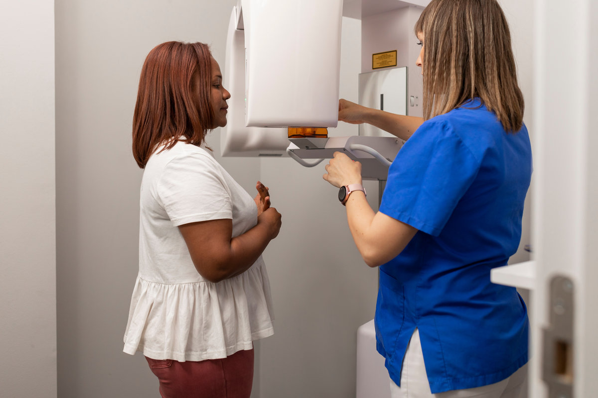
When tooth pain lingers well past what a normal sensitivity should, or when a previous root canal treatment stops feeling right, the questions come quickly: Is there an infection? Did something get missed? Is the tooth even savable? Standard dental X-rays have guided dentists for over a century, but they produce a flat, two-dimensional image of a three-dimensional structure. Bone loss hiding behind adjacent roots, fractures running along the wrong plane, extra canals tucked beneath calcified tissue, none of these reliably appear on a conventional radiograph. That gap between what a flat image shows and what is actually happening inside a tooth is exactly where patients get misdiagnosed, retreatment plans go sideways, and teeth that could have been saved get extracted.
At Burien Endodontics, Dr. Matthew Tomala uses cone-beam computed tomography (CBCT) as part of a diagnostic approach to see what flat films cannot. CBCT technology delivers a full three-dimensional image of the tooth, surrounding bone, and adjacent structures, providing an endodontist with the anatomical clarity that can change treatment decisions. If you have been told you need a root canal, are considering endodontic retreatment, or are dealing with persistent pain after a prior procedure, understanding how CBCT scanning works may help you ask better questions at your next appointment.
What a CBCT Scan Actually Shows
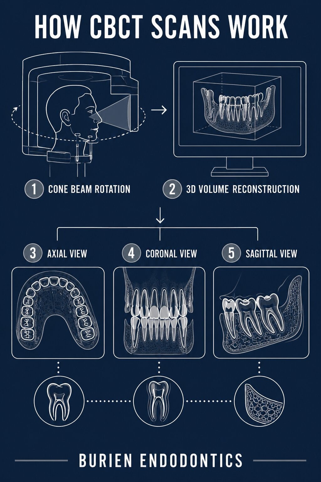 A CBCT scan works by rotating a cone-shaped X-ray beam around the head and capturing hundreds of images that software reconstructs into a three-dimensional volume. The endodontist can then navigate this volume in any plane: axial slices from above, coronal slices from the front, sagittal slices from the side. No single viewing angle is forced on the diagnosis. Every millimeter of root structure, canal anatomy, and surrounding bone becomes accessible from any direction.
This matters in endodontics because tooth anatomy is rarely as simple as a single root with a single canal. A 2025 evidence-based review in the clinical literature confirmed that CBCT offers better diagnostic accuracy across multiple endodontic applications than conventional radiography, including identifying complex anatomy, detecting apical periodontitis, and diagnosing root fractures. In practical terms, a tooth that appears to have two canals on a periapical film may reveal a third or fourth canal only visible in cross-section. Finding and treating all canals is the difference between a successful root canal and an unresolved infection.
Bone Loss and Periapical Lesions
One of the most clinically significant uses of CBCT is detecting periapical lesions, the areas of bone destruction that form around an infected root tip. These lesions are often present long before they show up on a two-dimensional X-ray, because a flat film only detects bone loss once it has progressed far enough to change the density visible from one specific angle. CBCT eliminates that blind spot. An endodontist can measure the true three-dimensional extent of a lesion, determine whether it is contained or spreading, and use that information to decide between root canal therapy and a surgical approach.
Root Fractures
Vertical root fractures are among the most difficult findings to confirm with conventional imaging. A fracture that runs along the long axis of a root may be essentially invisible on a periapical film because it lies parallel to the X-ray beam. CBCT scans can image the root from multiple angles simultaneously, making vertical fractures far more detectable. This is especially important before committing to retreatment: a fractured root often means the tooth cannot be saved regardless of how thorough the retreatment is, and identifying this up front protects the patient from an unnecessary procedure.
When CBCT Scanning Changes the Treatment Plan
Not every root canal case requires CBCT imaging, and responsible endodontic practice means calibrating diagnostic tools to clinical need. CBCT is most valuable when the case presents complexity that a standard radiograph cannot resolve. Some of the most common situations where CBCT scanning shifts the treatment plan include:
- A tooth with persistent or recurring pain after a completed root canal, where the source of failure is not visible on flat films
- Pre-surgical planning for an apicoectomy, where proximity to nerves or the sinus floor needs to be mapped before incision
- Suspected vertical root fracture, where the treatment decision changes completely depending on whether a fracture is confirmed
- Complex root canal anatomy, including C-shaped canals, fused roots, or unusual curvatures that could not be anticipated from a two-dimensional image
- Traumatic dental injuries, where the extent of root damage, bone involvement, or displacement needs to be assessed in three dimensions
In each of these situations, the CBCT scan does not simply produce a prettier picture. It answers clinical questions that would otherwise remain open. That certainty translates directly into a more accurate diagnosis, a more targeted treatment plan, and a better-informed conversation with the patient.
Learn More About Advanced Endodontic Diagnosis at Burien Endodontics
Burien Endodontics brings together CBCT scanning, Global A-Series microscopy, digital radiography, and laser technology under one roof, paired with Dr. Matthew Tomala’s postgraduate training and academic background. Whether you are facing a first root canal, a second opinion on a retreatment recommendation, or an endodontic surgery consultation, the diagnostic tools available here are built to give you clear answers before any treatment begins.
If you have questions about whether CBCT imaging is appropriate for your case, or if you would like to schedule a consultation with Dr. Tomala, please contact our office to get started.